 The average person breaks an estimated two bones in their lifetime. While broken bones are common injuries experienced across all ages, older individuals are at higher risk. Bones become more fragile and porous with age, so less force is needed to break their structure.
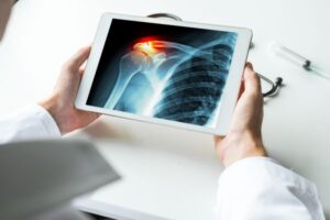
Medical imaging is used to help diagnose these injuries, so doctors can propose appropriate treatment plans. While X-rays are typically utilized, an MRI or CT scan may be recommended.
What Are Broken Bones?
Fractures occur when the bone is hit with a force greater than its mass, resulting in one of four types:
- Displaced Fracture: The bone breaks in at least two areas
- Non-Displaced Fracture: Characterized by a full or partial crack
- Closed Fracture: Indicates a break but no trauma to the skin
- Open Fracture: The bone breaks through the skin
Broken bones can quickly lead to complications based on severity, including infection and damage to blood vessels, nerves and surrounding tissue. Medical imaging assists in determining the extent of the damage.
Recovery may take a few weeks or months based on the type of break, the patient’s age and these potential complications.
Diagnosing a Broken Bone
Fractures almost always require emergency treatment. To determine if the bone is broken, doctors may use one of the following imaging technologies.
X-Ray Technology
A physical examination and X-ray results can help diagnose these injuries, especially in an emergency situation. Using a small amount of ionizing radiation, pictures of the body’s bones are captured.
This process delivers a quick, efficient approach for determining the presence of a fracture and if joint dislocation occurred. To allow for clear distinction, the imaging test will show the bones as nearly solid white and any surrounding tissue as gray.
CT Scans and MRIs
In certain instances, including for wrist, hip and stress fractures, an X-ray may not clearly indicate a break, despite the patient showing symptoms or experiencing pain. The doctor assessing your injury may recommend magnetic resonance imaging or a CT scan.
Ideal for assessing small fractures, the spinal cord, bone bruises and joint injuries, MRI technology provides more detail. This imaging test can indicate the presence of a broken bone, as well as a condition like a torn shoulder, ligament tear or rotator cuff injury. An MRI can also determine if the pain is coming from a microfracture or contusion.
A CT scan better indicates the presence of a fracture in patients with osteoporosis, as well as more complex fractures and skull fractures. This imaging test can also determine joint dislocation rather than a clean break.
Even if an X-ray shows a fracture, your doctor may recommend a CT scan or MRI for better visualization of damaged tissue and other secondary injuries.
Bone Scans
A bone scan is a type of nuclear imaging test that allows for better observation of certain bone diseases. Along with fractures, this procedure can indicate the presence of an infection, cancer or necrotizing bone tissue.
Doctors often order bone scans as a last resort, when a patient continues to experience pain and other signs of a bone condition, but other imaging technologies do not identify a break.
During this procedure, the patient is injected with a radioactive tracer, which travels where cells aid in repair. Medical imaging views the full skeleton to identify these spots and the condition present.
Along with identifying a fracture, bone scans can assist with diagnosing:
- Arthritis
- Cancer
- Bone and joint infections
- Paget’s disease
- Circulation issues
- Avascular necrosis, or dying bone tissue
Treatment for Fractures
Depending on the type and location of the fracture, treatment may involve:
- Cleaning the injury to prevent infection.
- Setting and positioning the bone in place. Surgery may be recommended with pins, screws or glue used to secure the bone.
- Joint replacement.
- Immobilizing the area with a cast, splint or traction device to help the bone heal.
- Medication, including painkillers and antibiotics.
- Physical therapy to improve motion and circulation, reduce atrophy and blood clots, and improve healing.
Midstate Radiology Associates can help diagnose a range of bone injuries. If your doctor has recommended an X-ray, MRI, CT scan or bone scan, schedule an appointment today.

 Accredited Facility Logo")


 Accredited Facility Logo")
 Accredited Facility Logo")

 Accredited Facility Logo")


