What Is a Breast Biopsy?
During a breast biopsy, a sample of tissue is removed for analysis. Imaging often guides the procedure to ensure the needle targets the precise location and provide a more accurate medical diagnosis.
There are two general types of biopsies. Fine needle aspiration uses a thin, hollow needle to extract fluid and a smaller portion of tissue, while a core needle biopsy removes a larger portion of tissue through a vacuum or spring-powered mechanism.
In select instances, a more invasive surgical biopsy may be requested. Ultrasound, MRI or mammography guides the needle to the exact location of the growth.
When Will a Doctor Request a Breast Biopsy?
If you recently had a mammogram, breast ultrasound or physical exam and the doctor noticed a lump or change in texture that could indicate cancer, a biopsy can investigate further. Common developments include:
- Thickening of breast tissue
- Nipple or areolar discharge
- Dimpling or scaling of the skin
- A lump or other growth felt on the breast or observed through imaging
- Possible calcium deposits seen in the breast tissue
- The presence of a cyst
Types of Breast Biopsies
The location and type of abnormality influences the breast biopsy your doctor may recommend:
- Fine Needle Aspiration: If your doctor felt a physical lump during a breast exam, you may be steered toward this type of biopsy, which involves collecting a sample of fluid or cells from the growth. Yet if its solid rather than filled with fluid, you may need to undergo a core needle biopsy to obtain a larger tissue sample.
- Core Needle Biopsy: Doctors often recommend a core needle biopsy if the lump is solid rather than fluid-filled. A hollow needle is used to extract a tissue sample, with ultrasound or MRI technology to guide the abnormality’s precise location.
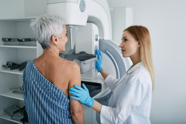
- Stereotactic Biopsy: Mammography guides this procedure, in which the breast is compressed. A small incision is made to extract multiple tissue samples.
- Surgical Biopsy: This more invasive procedure involves removing a larger portion of breast tissue. As such, patients are sedated or given local anesthetic. Guidance via wire, tag, seed and/or radioactive medicine may be used if the lump cannot be easily felt. Analysis includes establishing the presence of cancer and how far it has spread to surrounding tissue. Patients may undergo surgery to remove all tissue containing cancer cells.
Although these procedures are requested to rule out cancer, only 20 percent come back positive. In the remaining 80 percent of cases, the abnormality is a benign growth like a tumor, cyst or denser breast tissue.
What to Expect
Prior to undergoing a breast biopsy, discuss any allergies or blood thinners you take, if you have an implanted device or may be pregnant. This information helps your doctor determine whether to recommend a biopsy guided by ultrasound, MRI or mammography.
Ahead of the procedure:
- Understand that multiple biopsies may be needed to obtain a sufficient sample.
- Expect to be given local anesthetic unless a surgical biopsy is requested.
- Understand that a marker or clip may be added to the biopsy area to help a surgeon better identify the location of the growth, in the event more tissue needs to be removed in the future.
- Prepare to rest for the remainder of the day. With most needle-guided biopsies, you can typically go back to your normal routine within 24 hours.
- As bruising and tenderness may follow the procedure, you may be given acetaminophen and told to apply ice to the area to reduce any swelling.
Afterwards, look for signs of infection, bleeding or an altered appearance at the biopsy site. Aside from swelling, this can include fever, a warm sensation around the incision or abnormal drainage. If you notice any of these indicators, seek medical care immediately.
You should receive results within a few days once a pathologist analyzes the breast tissue and reports their findings to your doctor. Discrepancies may exist, such as imaging that shows signs of a precancerous condition, but a tissue analysis may come back with negative results. At this point, your doctor may request a more invasive procedure to obtain a larger tissue sample for analysis.
Has your doctor recommended a breast biopsy? Schedule your imaging procedure with Midstate Radiology Associates today.

